Latest News
GP Networking Day at the UCD School of Medicine
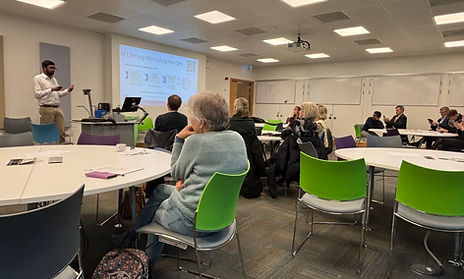
Dr. Alin Navas presented at the GP Networking Day at the UCD School of Medicine to introduce and promote the upcoming AI Literacy Workshop taking place on January 31, 2026. During the session, Dr. Navas highlighted the growing role of artificial intelligence in modern clinical practice and outlined the workshop’s purpose: to help clinicians better understand and critically evaluate the different types of AI systems now appearing in healthcare settings.
The workshop is designed to equip doctors with practical knowledge on how AI tools function, their limitations, and how to assess their safety, trustworthiness, and clinical relevance. Attendees at the networking day learned how this training aims to support GPs in integrating AI responsibly and confidently into patient care, ensuring informed decision-making as these technologies become more embedded in everyday workflows. Register at https://aiforgp.eventbrite.com/ .


Congratulations Dr. Belton !

Our posters at MIUA 2025
Yannuo Wen and Francesco Chiumento presented their papers at MIUA 2025.
Congratulations Dr. Bartels !


See you at ISBI 2025
Our research intern Nicolas Hadjittoouli has been contributing to the SMASH HCM project and is presenting his work at ISBI today. We're excited to share that Dr Atilla Krilly, from Google Health, expressed interest in his research.
Poster: Skull fractures, especially those of the base and facial regions, are challenging to identify due to the intricate anatomy and their subtle morphological characteristics. They necessitate meticulous attention and allocation of sufficient time to ensure accurate diagnosis and optimal patient care. This approach converts head CT to a mesh representation to utilize disk harmonic mapping and eventually create a flattened map of the skull for improved visualization. In such a view, fractures appear continuous with enhanced contrast. This alternative visualization of a flattened skull can provide radiologists and emergency department personnel with a valuable addition to conventional radiological views, increasing the effectiveness and speed of skull fracture detection.

Congratulations Dr. Belton !


Paper at NeurIPS 2024
Weichen Huang, a student at St. Andrew’s College and a recent intern at the lab, has won one of the top four awards in the first NeurIPS 2024 High School Track, a global competition highlighting impactful machine learning projects by students.
Weichen’s project, Multimodal Representation Learning Using Adaptive Graph Construction, focuses on improving Alzheimer’s disease detection. The method uses data from multiple sources, like images and text, to train AI models more effectively. The approach is not only more adaptable but also outperforms previous techniques, showing real potential for medical applications.
The NeurIPS High School Track, launched this year, received 330 submissions worldwide, with just 21 projects chosen for recognition. Weichen’s work stood out for its innovation and social impact. The winners presented their projects during a poster session on December 10, part of the prestigious NeurIPS 2024 conference.

.jpeg)

Carles’s PhD Graduation Celebration

.jpg)
Misgina’s PhD Graduation Celebration
UCD MIML Group celebrated Misgina’s PhD graduation and other ML-Labs graduates. We are sad to lose Misgina from our group and wish him the very best in his postdoctoral position in Linköping University, Sweden!

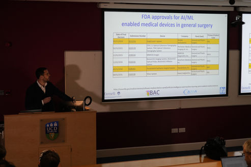
How AI is Shaping the Future of Surgery


Best Paper Award at MICAD 2024
Congrats to Yannuo Wen ! Best paper award to his (middle in the following picture) ‘Partition-based Medical Data Synthesis via Latent Diffusion’

Dr. Kathleen Curran’s Group at MICCAI 2024 in Marrakesh
Last week, members of Dr. Kathleen Curran’s group at UCD MLMI participated in the prestigious MICCAI 2024 conference held in Marrakesh, showcasing their latest research in medical imaging and machine learning.
Fangyijie Wang presented his work at the PIPPI workshop, where he introduced a novel approach using generative models to create synthetic fetal ultrasound images. His research is aimed at enhancing zero-shot classification for African cohorts, a significant step towards addressing underrepresented datasets in medical AI.
Additionally, Siteng Ma’s poster, titled "Adaptive Curriculum Query Strategy for Active Learning in Medical Image Classification," was presented at the conference. Her work explores new strategies to improve the efficiency and accuracy of medical image classification using active learning.
These presentations highlight the cutting-edge research being conducted by Dr. Curran's group and their ongoing contributions to the field of medical imaging and AI.
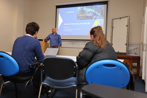
10+ Coding Workshop

Exciting Research Presentations by Fangyijie and Siteng at MICCAI 2024 !
We are thrilled to announce that Fangyijie and Siteng will be presenting our latest research findings at the prestigious MICCAI 2024 conference! As key contributors to our team's groundbreaking work in medical image analysis, they are eager to showcase two papers that delve into innovative techniques for advancing healthcare through artificial intelligence.
Fangyijie and Siteng are excited to engage with fellow researchers, healthcare professionals, and AI enthusiasts at MICCAI, discussing the potential impact of AI-driven solutions on medical practices worldwide.
Don't miss this opportunity to witness the future of medical imaging firsthand! Join us at MICCAI 2024 and support Fangyijie and Siteng as they present our team's latest innovations !
Best Paper Award at IMVIP 2024
Our student, Nighat Bibi, has won the Jon Campbell Best Paper Award at the IMVIP 2024, showcasing exceptional research and dedication. Congratulations to Nighat on this remarkable achievement!
Title: Enhancing Multiple Sclerosis Diagnosis with eXplainable AI


Two Papers accepted by MICCAI 2024
We are thrilled to announce that our paper has been accepted by MICCAI 2024 !
Title: Adaptive Curriculum Query Strategy for Active Learning in Medical Image Classification
Presenter: Siteng Ma
Overview: Active learning (AL) aims to reduce labeling costs by selecting informative samples for labeling. However, current AL methods often use a single query strategy throughout, which doesn't align with the learning process of deep learning models that first learn simple patterns before tackling complex ones. We propose an Adaptive Curriculum Query Strategy that adaptively changes the query strategy: initially selecting broadly distributed samples, and later shifting to an uncertainty-based strategy to focus on hard samples. Our method, validated with various diversity and uncertainty strategies, consistently improves performance.
We are thrilled to announce that we have one paper accepted for an oral presentation at MICCAI 2024 workshop PIPPI (Oral Presentation) !
Title: Generative Diffusion Model Bootstraps Zero-shot Classification of Fetal Ultrasound Images In Underrepresented African Populations
Workshop: Perinatal, Preterm and Paediatric Image Analysis (PIPPI)
Presenter: Fangyijie Wang
Overview: Developing robust deep learning models for fetal ultrasound image analysis requires comprehensive, high-quality datasets to effectively learn informative data representations within the domain. However, the scarcity of labelled ultrasound images poses substantial challenges, especially in low-resource settings. To tackle this challenge, we leverage synthetic data to enhance the generalizability of deep learning models. This study proposes a diffusion-based method, Fetal Ultrasound LoRA (FU-LoRA), which involves fine-tuning latent diffusion models using the LoRA technique to generate synthetic fetal ultrasound images. These synthetic images are integrated into a hybrid dataset that combines real-world and synthetic images to improve the performance of zero-shot classifiers in low-resource settings. Our experimental results on fetal ultrasound images from African cohorts demonstrate that FU-LoRA outperforms the baseline method by a 13.73% increase in zero-shot classification accuracy.
Accepted Paper at European Respiratory Society (ERS) 2024
Delighted to be accepted to present our work isolating the presence of Diffuse Cystic Lung Diseases with the RareLungAI group in Vienna. In this study, we manually segmented cysts in 22 high-resolution chest CTs from clinically confirmed Lymphangioleiomyomatosis cases with varying scan quality and disease burden.
Interpretability != Explainability
Dr. Gennady Roshchupkin is giving us a talk about XAI next week. Please join this session online via Zoom.

Oral Presentation at ATS 2024
Delighted to be accepted and present our work identifying the presence of Diffuse Cystic Lung Diseases (DCLDs) with the RareLungAI group at ATS (American Thoracic Society) International Conference 2024 in San Diego. In this study, 282 anonymised lung CTs with clinically confirmed diagnoses were used to train a deep-learning pipeline to identify DCLDs against Emphysema (a visually similar but more common condition) and "Normal" scans. During our trip, we also gathered valuable feedback from clinicians on our research and platform developed to screen for these diseases. Alongside this work, Evelyn Lynn also presented her research which analysed the use of semi-automated cyst volume assessments in establishing disease severity in DCLDs using our quantification pipeline.
Oral Presentation at BMFMS 2024
Dr. Helena Bartels was invited to present her research work at the recent British Maternal and Fetal Medicine Society’s (BMFMS) Annual Conference 2024. Her talk is about multi-centre study of antenatal prediction of FIGO grade 3 placenta accreta spectrum from multivariate T2-weighted MR imaging data.
Research Collaborations with Aix-Marseille University
Misgina Tsighe Hagos was awarded the France Excellence Research Residency 2024 grant for a research visit to the Methods and Computational Anatomy (MeCA) research group, Institut de Neurosciences de la Timone (INT) at Aix-Marseille University (AMU). Misgina will be working on brain imaging and preprocessing techniques for cortical thickness estimation and morphology of sulci and gyri.
Spring Symposium 2024

Dr. Ye Zi will give us a talk this seminar. Join us !
Please join us to welcome Dr. Ye to give a presentation about her recent research !
About Dr. Ye: Dr.Ye hold a PhD in Information and Communication Technology, specializing in computer vision and AI-driven medical imaging recognition. In her current role as a Postdoctoral Researcher at the Institute of Intelligent Software, she is at the forefront of developing AI applications for medical imaging. Her academic journey is enriched with significant teaching experience and leadership in AI-focused projects, including successful collaborations with industry partners. She has led and contributed to multiple high-impact research initiatives, resulting in a strong portfolio of publications in prestigious journals and conferences, such as IEEE Transactions on Geoscience and Remote Sensing (TGRS), International Conference on Image Processing (ICIP), and International Conference on Pattern Recognition (ICPR).


Winter Symposium 2024

Our recent work presented at NeurIPS 2023
PhD student, Niamh Belton attends the workshop ‘Medical Imaging meets NeurIPS’ at the Thirty-Seventh Annual Conference and Workshop on Neural Information Processing Systems in New Orleans to present her preliminary work on Rethinking Knee Osteoarthritis Severity Grading: A Few Shot Self-Supervised Contrastive Learning Approach. In this study, a continuous knee OA grading system is proposed to replace the current discrete Kellgren-Lawrence (KL) grading system. Self-supervised pre-training is used to learn a robust representation of healthy knee X-rays and the severity of OA is assessed based on the distance to the learned normal representation space.
PhD student, Misgina Tsighe Hagos presents his papers on a Distance-aware explanation based learning and a Postpartum family planning dataset from Addis Ababa, Ethiopia at the 7th BAI workshop at NeurIPS 2023.
Congratulations Niamh Belton and Misgina Tsighe Hagos !


MICCAI23 - Cardiac MRI reconstruction from undersampled k-space with the CMRxRecon challenge dataset
Julia Dietlmeier, Anam Hashmi, Carles Garcia-Cabrera, Kathleen M. Curran, Noel E. O'Connor
Abstract: The long acquisition time in cardiac MRI remains a major weakness of this diagnostic imaging approach. The undersampling of k-space by different factors (also known as the compressed sensing (CS) approach) offers accelerated data collection providing discomfort relief for paediatric patients. In addition to the motion artifacts inherently present in cardiac MRI, the process of frequency domain undersampling results in the loss of high-frequency information, which translates to a variety of noise and structural aliasing artifacts in the spatial domain, and thus further deteriorates the reconstructed image quality. In this work, we approach the problem of cardiac MRI image reconstruction from undersampled k-space using the newly released CMRxRecon dataset. Undersampled cardiac MRI is an inherently ill-posed problem leading to a variety of noise and aliasing artifacts if not appropriately addressed. We propose a two-step double-stream processing pipeline that first reconstructs a noisy sample from the undersampled k-space (frequency domain) using the inverse Fourier transform. Second, in the spatial domain we train a denoising GNA-UNET (enhanced by Group Normalization and Attention layers) on the noisy aliased and fully sampled image data using the Mean Square Error loss function. We achieve competitive results on the leaderboard and show that the algorithmic combination proposed is effective in high-quality MRI reconstruction from undersampled cardiac long-axis and short-axis complex k-space data.
This work has been a part of the CMRxRecon challenge hosted at the MICCAI 2023 conference in Vancouver, Canada. The challenge attracted 200 teams from 22 countries. We finished 15 on the leaderboard for the Task 1: cine reconstruction.


IMVIP23 - 25th Irish Machine Vision and Image Processing Conference, Galway
During my Transition Year work experience, I had the privilege of collaborating with researchers and students in the Machine Learning for Medical Imaging group, led by Dr. Kathleen Curran. Our collaboration kicked off in December 2022 with a symposium aimed at introducing pre-collegiate students in St Andrew's College from diverse backgrounds to the research carried out by our team. The primary goal was to ignite and nurture their interests in the fields of science and technology. The December symposium was a resounding success, achieving its goal of inspiring and engaging students. It showcased the power of effective collaboration between our research group and young, enthusiastic minds.
After the symposium, I joined the research group as an intern and became involved in lots of different projects, spanning topics such as explainable AI, image fusion, and Alzheimer's disease classification. Throughout this journey, I discovered the transformative potential of teamwork and the ability to learn from failures. In fact, most of my initial ideas didn't yield the promising results, but these failures ultimately led to valuable and crucial insights that I did not know previously.
One of the most impactful aspects of my experience was learning through presentation. With Dr. Kathleen Curran's guidance and support, I not only contributed to ongoing projects but also explored and addressed issues related to spurious correlations and data relevance in medical imaging. The pinnacle of this journey was when I proposed a solution and successfully presented our findings in a paper at the prestigious 25th Irish Machine Vision and Image Processing Conference.
During the conference, I gained lots of insight from other researchers who came from places all over Ireland. By asking questions, I was able to deepen my understanding of their research topics and methodologies, which sparked new and interesting ideas.
This experience was unlike anything I had ever encountered in my academic journey before, and I am extremely grateful for the opportunities and knowledge gained during this journey and look forward to continuing my pursuit of excellence in computer science and AI.
— Weichen, Huang
Accepted paper titled to Visual Anomaly and Novelty Detection Workshop at CVPR
Accepted paper titled 'FewSOME: One-Class Few Shot Anomaly Detection with Siamese Networks' to Visual Anomaly and Novelty Detection Workshop at CVPR with co-authors Misgina Tsighe Hagos, Aonghus Lawlor and Kathleen Curran.
Awarded a Short Term Scientific Mission (STSM) from Genomics of MusculoSkeletal traits Translational Network (GEMSTONE)
Awarded a Short Term Scientific Mission (STSM) from Genomics of MusculoSkeletal traits Translational Network (GEMSTONE), funded by the EU, to travel to Erasmus University Rotterdam to work on a European Cooperation in Science and Technology (COST) action that will involve using anomaly detection techniques to identify poor quality images.
Machine Learning to Rule-Out Pulmonary Embolism
Congratulations to Dr Nicholas McCarthy for his recent "Extended D-dimer Cut-offs and Machine Learning for Ruling Out Pulmonary Embolism in individuals undergoing CTPA" publications in the European Respiratory Journal March 2022. Checkout the publication here.

Research Collaborations between Ireland and France-based Researchers
In June 2021, Misgina Tsighe Hagos and Niamh Belton, under the supervision of Prof. Kathleen Curran, were granted Ulysses funding by the Irish Research Council (IRC). Ulysses funding aims to promote research collaborations between Ireland and France-based researchers. We are using the funding to collaborate with the Center for Magnetic Resonance in Biology and Medicine (CRMBM) research group, based in Aix-Marseille. This is a research group that specialises in musculoskeletal medical imaging and have acquired datasets of Magnetic Resonance Imaging (MRI) and Diffusion Tensor Imaging (DTI). These datasets are valuable as measurements of the muscle architecture can be more accurately calculated in 3D space from DTI. Such measurements can provide critical information on injury risk, injury recovery process, and muscle disorders. Given our skills in Machine Learning and their expertise in musculoskeletal imaging, this collaboration facilitates an exciting opportunity to develop novel machine learning models for automating muscle architecture analysis.
As part of the collaboration, we had the pleasure of hosting Prof. David Bendahan, CNRS research director and leader of the “Magnetic Resonance MSK” group at CRMBM, on a research visit for three days where we exchanged research ideas in November 2021. He also gave an interesting seminar on ‘Segmentation of individual muscles in MR images: How could we handle pathological changes?’. Following this, we travelled to Marseille for four weeks where we met with various members of the research group. We began discussions to define the project and began curating datasets. We are looking forward to the next meeting when the members of CRMBM will travel to Ireland.
2021 Virtual Mobility Grant for “Differentiating Between Muscle and Fat Tissue in Zebrafish Using AI”
2021 Virtual Mobility Grant supported by the COST Action Strategy under the GEnomics of MusculoSkeletal traits Translational NEtwork (GEMSTONE), functional WG2 and WG4. Received under collaboration of Dr. David Karasik, Bar-Ilan University (Host Institution), and Dr. Kathleen Curran, University College Dublin (Home Institution). The grant was used to work on the project “Differentiating Between Muscle and Fat Tissue in Zebrafish Using AI”.
2021 Virtual Mobility Grant for “Accelerating Genetic Screening of Skeletal Conditions Using Zebrafish and AI”
2021 Virtual Mobility Grant supported by the COST Action Strategy under the GEnomics of MusculoSkeletal traits Translational NEtwork (GEMSTONE), functional WG4. Received under collaboration of Dr. Erika Kague, University of Bristol (Host Institution), and Dr. Kathleen Curran, University College Dublin (Home Institution). The grant was used to further work on the project “Accelerating Genetic Screening of Skeletal Conditions Using Zebrafish and AI”.
We are delighted to welcome our collaborator, Ms. Susan Maguire, to our research group
Ms. Susan Maguire is a Medical Physicist at the Mater Private Hospital. Ms. Maguire trained in Addenbrookes hospital, Cambridge in the IPEM training program and moved to the Mater Private 16 years ago where she is currently the Chief Physicist in Diagnostic Imaging. Ms. Maguire acts as the hospital's Radiation Protection Advisor and Medical Physics Expert. They cover a wide range of modalities including CT, PET/CT, nuclear medicine, interventional radiology/cardiology, x-ray, mammography as well as MRI and non-ionizing modalities such as lasers, UV, and ultrasound across 7 sites. Ms. Maguire currently lectures on the UCD MSc in Medical Physics course and has supervised a number of projects. Ms. Maguire is also vice president of the Irish College of Physicists in Medicine. The day-to-day work doesn't leave much time for research but she has collaborated with UCD on a number of projects and looks forward to more in the future.

We are delighted to welcome our collaborator, Dr Jim O'Doherty, to our research group
Dr O'Doherty is a clinical scientist by background, He trained in the UK medical physics program and spent most of my time over the last few years setting up and analyzing clinical trials (on both humans and animals) using MR, PET, CT, and hybrid systems like PET-MR, PET-CT. Dr O'Doherty also worked on the development of new radiotracers for imaging various pathways in cancer (mainly breast and thyroid). Dr O'Doherty does some research in detector physics through designing and simulating new detectors, especially small field of view PET systems, but also a newly released photon-counting CT system (Siemens). Dr O'Doherty is currently working in the USA as an R&D collaborations manager for Siemens, where they develop new imaging techniques and quite a lot of AI postprocessing software (including some at the image acquisition stage) along with their clinical collaborators. Dr O'Doherty has an academic post at the Medical University of South Carolina where he is based.

Congratulations to Yushi Yang on receiving VM grant for implementing AI to automate swimming behaviour analysis of zebrafish for musculoskeletal research
Yushi is collaborating with Kathleen Curran and Erika Kague to develop a deep learning model for tracking a large amount of fish, in order to study their 3D collective behaviour.
Zebrafish have been used in biomedical research to study gene function, identification of targets for therapeutics and to test drugs to modulate diseases. By studying the zebrafish swimming behaviour we are able to study how a gene can affect the musculoskeletal system and the zebrafish vertebral column mobility, similar to gait analysis in human. The key step for the behaviour study is to reconstruct the trajectories of animals. With a recent virtual mobility grant (COST Action: CA18139), Yushi Yang (Physics PhD student in Bristol) is further optimising a novel 3D fish tracking system, by applying deep learning models. The efficiency of the 3D tracking system is expected to get a tenfold increase, enabling fast genetic screenings.

Congratulations to Niamh Belton, Ivan Welaratne, Adil Dahlan, Ronan T Hearne, Misgina Tsighe Hagos, Aonghus Lawlor, and Kathleen M. Curran for receiving the best paper and presentation award at MIUA 2021 for their paper titled 'Optimising Knee Injury Detection with Spatial Attention and Validating Localisation Ability'
Linkk to the pp

Congratulations To Our Research Group Members: Niamh Belton and Misgina Tsighe Hagos Upon Receiving Ulyssess Funding
Niamh Belton and Misgina Tsighe Hagos, under the supervision of Prof. Kathleen Curran, Prof. Brian McNamee and Prof. Aonghus Lawlor, have been awarded Ulysses funding from the Irish Research Council to travel to France to collaborate with the CRMBM research group in Aix-Marseille University. The aim of the Ulysses scheme is to foster new collaborations between Ireland- and France-based researchers. The project will focus on using Machine Learning to automate the segmentation of muscles from Magnetic Resonance Imaging (MRI) and Diffusion Tensor Imaging (DTI) data.
Niamh Belton and Misgina Tsighe Hagos, under the supervision of Prof. Kathleen Curran, Prof. Brian McNamee and Prof. Aonghus Lawlor, have been awarded Ulysses funding from the Irish Research Council to travel to France to collaborate with the CRMBM research group in Aix-Marseille University. The aim of the Ulysses scheme is to foster new collaborations between Ireland- and France-based researchers. The project will focus on using Machine Learning to automate the segmentation of muscles from Magnetic Resonance Imaging (MRI) and Diffusion Tensor Imaging (DTI) data.

MIDL 2021 Publication
Congratulations to Niamh Belton, member of our research group on her latest publication with Dr Kathleen Curran and Aonghus Lawlor.
If you are interested, make sure to attend MIDL 2021, Friday the 9th session at 16:45 (UCT+2) to hear Niamh speak about the paper!

AI Enhanced DIffusion MRI in Neuroimaging
Recent advances in artificial intelligence provide a new era of opportunities for new methodologies to address challenges in diffusion MRI. Large open source labelled data sets and open source code for emerging frameworks provide opportunities to train diffusion MRI models to drive efficiencies in acquisition and reconstruction and augment clinical decision making. This is particularly pertinent in the case of interpretability of diffusion MRI, which is significantly more computationally expensive and challenging than conventional MRI in diagnosis and prediction of neurological disorders.
We encourage the submission of scientific papers (original research, reviews, perspectives, etc.) focused on machine learning and artificial intelligence with clinical applicability for diagnosis and prediction.
For more information:
TOPIC EDITORS
Kathleen Curran, University College Dublin, Ireland
Daan Christiaens, KU Leuven, Belgium
Eleftheria Panagiotaki, University College London, UK
Paddy Slator, Univeristy College London, UK

LATEST PUBLICATION!
Enterprise imaging and big data: A review from a medical physics perspective
Abstract: In recent years enterprise imaging (EI) solutions have become a core component of healthcare initiatives, while a simultaneous rise in big data has opened up a number of possibilities in how we can analyze and derive insights from large amounts of medical data. Together they afford us a range of opportunities that can transform healthcare in many fields. This paper provides a review of recent developments in EI and big data in the context of medical physics. It summarizes the key aspects of EI and big data in practice, with discussion and consideration of the steps necessary to implement an EI strategy. It examines the benefits that a healthcare service can achieve through the implementation of an EI solution by looking at it through the lenses of: compliance, improving patient care, maximizing revenue, optimizing workflows, and applications of artificial intelligence that support enterprise imaging. It also addresses some of the key challenges in enterprise imaging, with discussion and examples presented for those in systems integration, governance, and data security and privacy.

FUSION Exemplar Award 2019
In partnership with Axial Medical Printing Ltd. Dr Kathleen Curran and Mr John Fitzpatrick were awarded the 2019 InterTrade Ireland FUSION Project Exemplar Award. Mr Fitzpatrick is now a PhD student ML-Labs (cohort 1).


